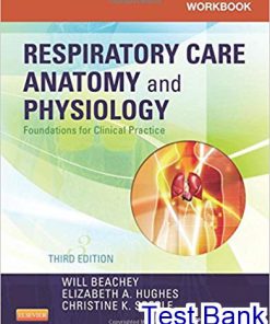
Respiratory Care Anatomy and Physiology 3rd Edition Beachey Test Bank
$50.00 Original price was: $50.00.$26.50Current price is: $26.50.
Respiratory Care Anatomy and Physiology 3rd Edition Beachey Test Bank.
Instant download Respiratory Care Anatomy and Physiology 3rd Edition Beachey Test Bank pdf docx epub after payment.
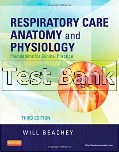
Product Details:
- ISBN-10 : 0323078664
- ISBN-13 : 978-0323078665
- Author: Will Beachey
Table of contents:
- Section I The Respiratory System
- Chapter 1 The Airways and Alveoli
- Objectives
- Key Terms
- The Airways
- Upper Airways
- Nose
- Figure 1-1 Structure of the respiratory system. Note the division between the upper and lower respiratory tracts.
- Figure 1-2 The bony nasal septum.
- Figure 1-3 The nasal cavity and pharynx viewed from the medial side.
- CONCEPT QUESTION 1-1
- Figure 1-4 An oral endotracheal tube in position. The inflated cuff at the tip of the tube isolates the lower trachea and airways from the pharynx.
- CONCEPT QUESTION 1-2
- Pharynx
- CONCEPT QUESTION 1-3
- Clinical Focus 1-1
- Endotracheal Tubes, Drying of Secretions, and Humidification Goals
- Discussion
- Figure 1-5 Head position affects airway patency. A, A flexed head may occlude the airway. B, The normal position of the head and neck. C, An extended head aligns the oral cavity and pharynx with the trachea, opening the airway and facilitating intubation.
- Larynx
- Figure 1-6 Anatomy of the larynx. A, Anterior view. B, Posterior view.
- Clinical Focus 1-2
- Treatment of Obstructive Sleep Apnea (OSA)
- Figure 1-7 Vocal cords, epiglottis, and glottis. A, Vocal cords viewed from above. B, Photographic view of the same structures.
- Clinical Focus 1-3
- Seriousness of Airway Edema in Adults and Young Children
- Discussion
- CONCEPT QUESTION 1-4
- Lower Airways
- Trachea and Main Bronchi
- Figure 1-8 The tracheobronchial tree. A plastic cast of airspaces in the human lung is shown.
- CONCEPT QUESTION 1-5
- Figure 1-9 The trachea and mainstem, lobar, and segmental bronchi. The extrapulmonary-intrapulmonary boundary marks the point at which the mainstem bronchi penetrate the lung tissue.
- Figure 1-10 Normal temperature and humidity of inspired gas during spontaneous breathing at different points along the respiratory tract. RH, Relative humidity.
- Conducting Airway Anatomy
- Figure 1-11 Branching of the conducting and terminal airways. Alveoli first appear in the respiratory bronchioles, marking the beginning of the respiratory or gas-exchange zone. BR, Bronchus; BL, bronchiole; TBL, terminal bronchiole; RBL, respiratory bronchiole; AD, alveolar duct; AS, alveolar space; Z, order of airway division.
- Clinical Focus 1-4
- Absent Breath Sounds after Intubation
- Discussion
- CONCEPT QUESTION 1-6
- Figure 1-12 Lobes and segments of the lungs.
- Clinical Focus 1-5
- Airway Anatomy and Drainage of Lung Secretions
- Discussion
- Sites of Airway Resistance
- CONCEPT QUESTION 1-7
- Conducting Airway Histology
- Figure 1-13 Gas-exchange portion of the lung. These subdivisions of the terminal bronchiole form the acinus.
- Figure 1-14 Distribution of airway cross-sectional area in the lung. The airways less than 2 mm in diameter collectively form a much larger cross-sectional area than the area formed by larger airways. Flow resistance of small airways is therefore less than flow resistance of large airways.
- Other Epithelial Cells
- Mucociliary Clearance Mechanism
- TABLE 1-1 Subdivisions of the Respiratory Tree
…
People Also Search:
Download full solution manual at: Respiratory Care Anatomy and Physiology 3rd Edition Beachey
Download full test bank at: Respiratory Care Anatomy and Physiology 3rd Edition Beachey
Download full solution manual + test bank at: Respiratory Care Anatomy and Physiology 3rd Edition Beachey
Download full test bank + solution manual at: Respiratory Care Anatomy and Physiology 3rd Edition Beachey
testbank download pdf Respiratory Care Anatomy and Physiology 3rd Edition Beachey
download scribd Respiratory Care Anatomy and Physiology 3rd Edition Beachey
solution manual download pdf Respiratory Care Anatomy and Physiology 3rd Edition Beachey
Instant download after Payment is complete
You may also like…
Anatomy and Physiology
Test Bank
Cardiopulmonary Anatomy Physiology Essentials of Respiratory Care 6th Edition Jardins Test Bank
Solutions Manual


