Ecgs Made Easy 5th Edition Aehlert Test Bank
$50.00 Original price was: $50.00.$26.50Current price is: $26.50.
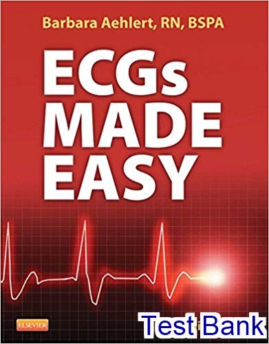
Ecgs Made Easy 5th Edition Aehlert Test Bank.
Categories: Other, Test Bank
Tags: Aehlert, Ecgs Made Easy, Test Bank
Instant download Ecgs Made Easy 5th Edition Aehlert Test Bank pdf docx epub after payment.
Product Details:
- ISBN-10 : 0323101089
- ISBN-13 : 978-0323101080
- Author: Barbara J Aehlert MSEd BSPA RN
Table of contents:
- CHAPTER 1 Anatomy and Physiology
- LEARNING OBJECTIVES
- KEY TERMS
- LOCATION, SIZE, AND SHAPE OF THE HEART
- [Objective 1]
- SURFACES OF THE HEART
- [Objective 2]
- Figure 1-1 Anterior view of the chest wall of a man showing skeletal structures and the surface projection of the heart.
- Figure 1-2 Appearance of the heart. This photograph shows a living human heart prepared for transplantation into a patient. Note its size relative to the hands that are holding it.
- Figure 1-3 The base of the heart.
- Figure 1-4 The anterior surface of the heart.
- COVERINGS OF THE HEART
- [Objective 3]
- CLINICAL CORRELATION
- Figure 1-5 The inferior surface of the heart. The inferior part of the fibrous pericardium has been removed with the diaphragm.
- CLINICAL CORRELATION
- STRUCTURE OF THE HEART
- Layers of the Heart Wall
- [Objective 4]
- Did You Know?
- Figure 1-6 The fibrous pericardium and phrenic nerves revealed after removal of the lungs.
- Figure 1-7 The fibrous pericardium has been opened to expose the visceral pericardium covering the anterior surface of the heart.
- Figure 1-8 Wall of the heart. The cutout section of the heart wall shows the outer fibrous pericardium and the parietal and visceral layers of the serous pericardium (with the pericardial space between them). Note that a layer of fatty connective tissue is located between the visceral layer of the serous pericardium (epicardium) and the myocardium. Note also that the endocardium covers beamlike projections of myocardial muscle tissue, called trabeculae carneae (meaning “fleshy beams”) that help to add force to the inward contraction of the heart wall.
- Table 1-1 Layers of the Heart Wall
- Cardiac Muscle
- Figure 1-9 A, Cardiac muscle fiber. Unlike other types of muscle fibers, the cardiac muscle fiber is typically branched and forms junctions, called intercalated disks, with adjacent cardiac muscle fibers. B, Thin myofilament. C, Thick myofilament.
- ECG Pearl
- Heart Chambers and Valves
- Atria
- [Objective 5]
- Figure 1-10 Interior of the heart. This illustration shows the heart as it would appear if it were cut along a frontal plane and opened like a book. The front portion of the heart lies to the reader’s right; the back portion of the heart lies to the reader’s left. (Note each portion has a separate anatomical rosette to facilitate orientation.) The four chambers of the heart—two atria and two ventricles—are easily seen. AV, atrioventricular; SL, semilunar.
- Figure 1-11 Section through the heart showing the apical portions of the left and right ventricles.
- ECG Pearl
- Ventricles
- [Objective 5]
- CLINICAL CORRELATION
- Heart Valves
- Figure 1-12 Skeleton of the heart. This posterior view shows part of the ventricular myocardium with the heart valves still attached. The rim of each heart valve is supported by a fibrous structure, called the skeleton of the heart, which encircles all four valves. AV, Atrioventricular.
- Figure 1-13 Internal view of the right ventricle.
- Atrioventricular Valves
- [Objectives 6, 7]
- Figure 1-14 Internal view of the left ventricle.
- Semilunar Valves
- [Objective 6]
- CLINICAL CORRELATION
- Heart Sounds
- Figure 1-15 Drawing of a heart split perpendicular to the interventricular septum to illustrate the anatomic relationships of the leaflets of the atrioventricular and aortic valves.
- CLINICAL CORRELATION
- The Heart’s Blood Supply
- Coronary Arteries
…
People Also Search:
Download full solution manual at: Ecgs Made Easy 5th Edition Aehlert
Download full test bank at: Ecgs Made Easy 5th Edition Aehlert
Download full solution manual + test bank at: Ecgs Made Easy 5th Edition Aehlert
Download full test bank + solution manual at: Ecgs Made Easy 5th Edition Aehlert
testbank download pdf Ecgs Made Easy 5th Edition Aehlert
download scribd Ecgs Made Easy 5th Edition Aehlert
solution manual download pdf Ecgs Made Easy 5th Edition Aehlert
Instant download after Payment is complete
You may also like…
Sale!
Test Bank
Sale!
Solutions Manual
Sale!
Management
Sale!
Sale!
Calculus and Mathematics
Sale!
Sale!
Test Bank
Sale!
Sale!
Related products
Sale!
Sale!
Sale!
Sale!
Sale!
Sale!
Sale!
Sale!


